Diagnostic imaging
Diagnostic imaging for orthopedic conditions
When you experience an injury or illness, you want to find answers as soon as possible. At TRIA, we use the latest orthopedic imaging technologies to accurately diagnose your condition and begin working on a personalized treatment plan.
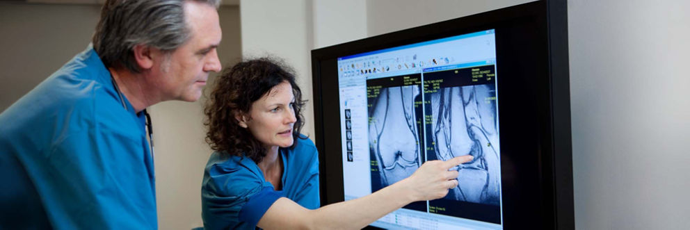
Our diagnostic imaging services include digital MRI (magnetic resonance imaging), X-rays, C-arms and ultrasound. In addition, TRIA has a musculoskeletal radiologist and neuroradiologist on staff, who are experts in interpreting diagnostic imaging.
Get started by making an appointment for diagnostic imaging services, and together we’ll get you back to your active lifestyle.
Diagnostic imaging services we provide
Our fully digital diagnostic imaging equipment gives us accurate and timely information about the location, nature and scope of an injury. Our orthopedic imaging services include:
Magnetic Resonance Imaging (MRI)
An MRI uses magnetic energy to create a detailed picture of the soft tissue within your body. During an MRI, you’ll lie on your back inside a tube while the digital images are acquired.
We use the latest digital imaging equipment from the leaders in MRI technology. This includes high-definition MRI scanners that allow doctors to more easily diagnose even the most challenging cases.
We also have one short, wide-bore MRI machine that patients often find more comfortable because more of the body is outside the bore, and there’s more room around the patient. Not only does this provide shorter scanning times, it helps people with claustrophobia to complete their exams.
Digital X-rays
An X-ray uses energy waves to take a picture of your bones. Some doctors take the picture on a film that needs time to develop. At TRIA, we use digital X-rays.
Rather than waiting for films to be developed, digital X-rays are transmitted immediately to be viewed by doctors – and there’s no waiting period. This means we can more quickly diagnose bone disorders and recommend a treatment plan.
C-arm imaging
A C-arm is an imaging scanner intensifier. It’s called a C-arm because of its “C” shape, and because it can be connected to a digital X-ray to provide high-resolution images in real time.
Thanks to its shape, the C-arm can take fluoroscopic images of the body from virtually any angle, side or position. During this scan, patients don’t need to move – the C-arm allows them to remain in one place while the image is taken.
Musculoskeletal ultrasound imaging
An ultrasound, sometimes called a sonogram, uses soundwaves to create a picture of the inside of your body. This allows us to examine your tendons and ligaments, and check for foreign bodies or fluid collections.
We can also use an ultrasound to guide us during treatments. For example, an ultrasound often helps us administer injections, especially when we’re treating ganglion cysts and tendon issues.
A dynamic ultrasound, which is a type of ultrasound performed while the patient moves the part of the body being examined, can be used to check for ligament instabilities, tendon injuries and dislocations.
